
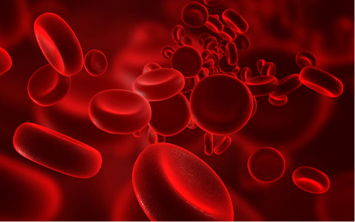
Hemolyzed blood samples are one of the most common reasons laboratories reject and repeat tests. Hemolysis occurs when red blood cells rupture and release hemoglobin and intracellular contents into the serum or plasma. This process can significantly alter test results for potassium, AST, LDH, and many other analytes, leading to misinterpretation and delayed diagnosis. Both preanalytical errors and patient-related factors can cause hemolysis, so clinicians and phlebotomists must understand where problems start. By recognizing high‑risk steps during blood collection, handling, and transport, healthcare teams can reduce sample rejection rates and improve the reliability of laboratory data.
Common Causes of Hemolyzed Blood in Laboratory Samples
Most hemolyzed blood samples result from preanalytical issues such as poor venipuncture technique, wrong equipment, improper mixing, and rough transport conditions that physically damage red blood cells before testing.
Improper Blood Collection Techniques and Equipment Issues
Poor venipuncture technique is a leading cause of hemolysis. Using a needle that is too small increases shear stress and ruptures red blood cells as blood enters the tube. Pulling back hard on a syringe plunger or forcing blood through a needle into a tube also causes mechanical damage. Prolonged tourniquet application and excessive fist pumping raise pressure in the vein and promote cell rupture. Incorrect order of draw and underfilling tubes with liquid anticoagulant alter blood-to-additive ratios. Using damaged needles, faulty collection sets, or expired tubes further increases hemolysis risk and leads to unreliable lab results.
Sample Handling and Transportation Errors in the Preanalytical Phase
Even when the collection is perfect, poor handling can still produce hemolyzed samples. Vigorous shaking of tubes rather than gentle inversion damages red blood cells. Delays in separating serum or plasma from cells allow ongoing metabolic changes and increase the chance of hemolysis. Transporting specimens in pneumatic tube systems at high speeds or with sudden stops adds mechanical shock. Exposure to extreme temperatures during storage or transport also weakens red cell membranes. Failure to follow recommended mixing times for anticoagulant tubes or transporting uncapped tubes increases both hemolysis and contamination risks, compromising the reliability of subsequent test results.
Environmental and Mechanical Factors That Damage Red Blood Cells
Red blood cells are sensitive to physical and environmental stress. Extreme heat or cold during storage can destabilize cell membranes and trigger hemolysis. Freezing whole blood that is not intended for freezing causes ice crystal formation and cell rupture. High‑speed centrifugation, using incorrect rotor settings, or spinning samples for too long, places excessive force on cells. Leaving samples exposed to sunlight or near heat sources accelerates degradation. Vibrations from transport equipment, shaking coolers, or long trips over rough surfaces can also damage cells. These environmental and mechanical factors often combine with collection errors to produce markedly hemolyzed specimens.

Patient-Related and Biological Factors Contributing to Hemolysis
Some patients have inherently fragile red blood cells or ongoing intravascular hemolysis. These biological factors can cause hemolyzed samples even when phlebotomy, handling, and transport follow best laboratory practices.
Underlying Medical Conditions and Red Blood Cell Fragility
Certain clinical conditions make red blood cells more prone to rupture. Hereditary spherocytosis, G6PD deficiency, sickle cell disease, and other hemoglobinopathies reduce membrane stability and shorten red cell survival. Autoimmune hemolytic anemia and transfusion reactions cause immune‑mediated destruction of red blood cells. Severe infections, sepsis, burns, and disseminated intravascular coagulation can trigger widespread intravascular hemolysis. Patients on some drugs or with heavy metal exposure may develop toxic injury to red cells. In these cases, hemolysis can occur in vivo, and the resulting samples appear hemolyzed even when collection and handling strictly follow standard laboratory guidelines.
Physiological Stress, Dehydration, and Electrolyte Imbalances
Intense physical exertion, prolonged seizures, or severe physiologic stress can increase red blood cell breakdown in the circulation. Dehydration reduces plasma volume, concentrates blood, and increases viscosity, making cells more vulnerable to shear stress during venipuncture and circulation. Marked electrolyte disturbances, such as extreme hypo‑ or hypernatremia, alter osmotic balance across red cell membranes and promote cell swelling or shrinkage, which may end in rupture. Severe burns or crush injuries mechanically destroy red cells and release free hemoglobin. These conditions can produce samples with visible hemolysis and abnormal test results, complicating the interpretation of routine chemistry and hematology panels.
In Vivo vs In Vitro Hemolysis: Key Differences in Origin
In vivo hemolysis occurs inside the patient’s circulation before blood collection, usually due to disease, immune reactions, toxins, or severe physical stress. It often presents with clinical signs like anemia, jaundice, dark urine, and elevated LDH and bilirubin. In vitro hemolysis happens after blood leaves the body, typically from poor technique or handling. Patients usually have no clinical evidence of hemolytic anemia, and repeat samples collected correctly may appear normal. Distinguishing in vivo from in vitro hemolysis is essential because in vitro hemolysis mainly indicates preanalytical error, while in vivo hemolysis represents a true pathological process needing investigation.
Conclusion
Hemolyzed blood samples arise from a mix of technical, environmental, and patient‑related factors. Small needles, forceful collection, rough mixing, and improper transport remain the most frequent causes and are largely preventable through staff training and standard operating procedures. Environmental extremes and equipment misuse can further damage red cells and distort test results. At the same time, clinicians must recognize when hemolysis reflects true in vivo pathology rather than preanalytical error. Clear communication between phlebotomy, laboratory, and clinical teams, supported by consistent quality control, reduces hemolysis rates and ensures more accurate and reliable laboratory data for patient care.